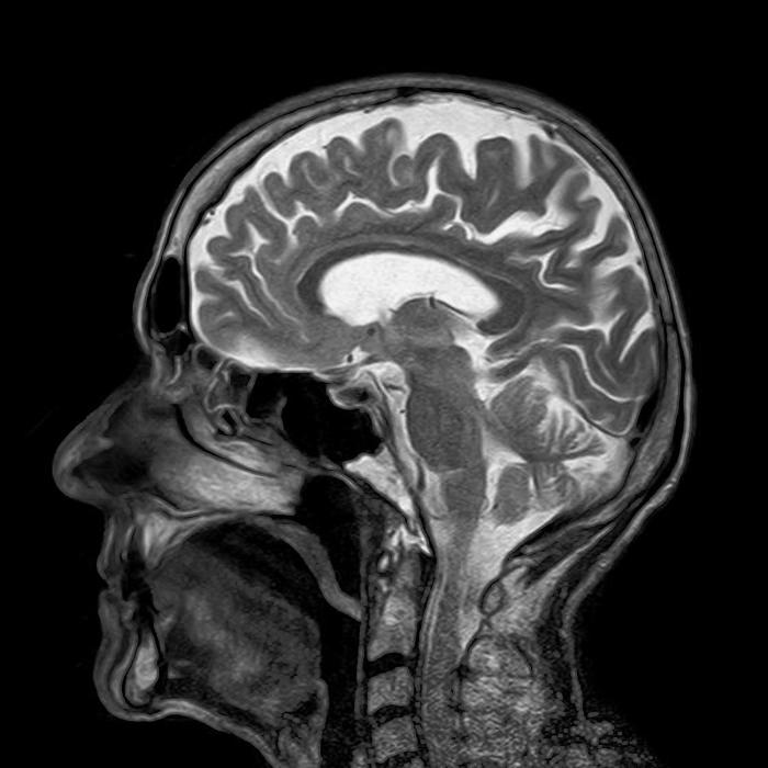
Faster mri scans ai machine learning facebook nyu research clinical study – Faster MRI scans: AI machine learning, Facebook, NYU research, clinical study. Imagine medical imaging so fast, it revolutionizes diagnosis and treatment. This groundbreaking research from Facebook and NYU is exploring how AI can dramatically speed up MRI scans, potentially saving valuable time and resources in clinical settings. The project leverages cutting-edge machine learning algorithms to accelerate the process while maintaining image quality, paving the way for a future where quicker and more accessible diagnostics are a reality.
This promises to significantly improve patient care by allowing doctors to get critical information faster, leading to better and quicker treatment decisions.
The study delves into the intricate process of AI-accelerated MRI scans, from data acquisition and preprocessing to the training and validation of AI models. This detailed approach ensures accuracy and reliability, crucial for clinical applications. Key considerations like ethical implications and the potential impact on healthcare systems are also examined. The collaboration between Facebook’s AI expertise and NYU’s medical imaging prowess underscores the potential of technology to transform healthcare.
Introduction to Faster MRI Scans
Magnetic Resonance Imaging (MRI) is a powerful diagnostic tool, providing detailed images of the body’s internal structures. However, standard MRI scans can be lengthy, often taking 30 minutes or more, which can be problematic for patients with claustrophobia, young children, or those with certain medical conditions. This prolonged scan time also impacts workflow in clinical settings. The need for faster MRI scans is clear.Current MRI technology relies on complex interactions between radio waves, magnetic fields, and the body’s tissues.
While high-resolution images are possible, the inherent nature of these interactions limits the speed at which data can be acquired. This limits the acquisition of high-quality images in a short timeframe. Accelerating MRI scans while maintaining image quality is a significant challenge.
Potential of AI and Machine Learning
Artificial intelligence (AI) and machine learning (ML) offer promising avenues for accelerating MRI scans. These techniques can analyze large datasets of MRI images to identify patterns and develop algorithms that predict and reconstruct missing data points. By effectively filling in the gaps, AI can shorten the scan time significantly without compromising image quality.
Role of Facebook and NYU
Facebook’s AI research division and New York University (NYU) have been actively collaborating on developing AI-powered MRI acceleration techniques. Facebook’s deep learning expertise combined with NYU’s medical imaging expertise creates a powerful synergy. This partnership leverages the strengths of both institutions to tackle the complexities of medical imaging.
General Approach to AI-Accelerated MRI Scans
The general approach involves training AI models on vast datasets of existing MRI scans. These models learn to predict the missing data points during the scan, enabling the reconstruction of a high-quality image from a significantly reduced amount of raw data. This approach involves training neural networks on a large set of MRI images to identify patterns in the data.
The resulting models can then predict the missing data points, thereby accelerating the scan process. A common strategy involves utilizing deep learning architectures, such as convolutional neural networks (CNNs), to process and interpret the image data.
Recent research from Facebook and NYU, using AI machine learning to speed up MRI scans in clinical studies, is pretty cool. This advancement could revolutionize how we diagnose and treat illnesses. Interestingly, this groundbreaking work is connected to a fascinating legal battle, where X is backing one of the smallest stakes legal fights imaginable. This seemingly disparate legal dispute actually highlights the complex interplay between innovation and regulation in the healthcare field.
The potential for faster MRI scans is still very exciting, and it’s fascinating to see how this technology will evolve.
Comparison of Scan Times
| Current MRI Scan Time | Proposed AI-Accelerated Scan Time | Potential Advantages |
|---|---|---|
| 30-60 minutes | 5-15 minutes | Reduced patient discomfort, improved patient workflow, and faster turnaround time for diagnosis. |
The table illustrates the potential reduction in scan time using AI-accelerated techniques. A significant reduction in scan time is possible, potentially leading to improved patient experiences and increased efficiency in clinical settings.
AI Algorithms for MRI Acceleration
The quest for faster magnetic resonance imaging (MRI) scans is a significant endeavor in medical imaging. Traditional MRI techniques can be time-consuming, limiting their use in certain clinical scenarios, particularly in emergency situations or for patients with claustrophobia. Artificial intelligence (AI) algorithms offer a promising avenue to accelerate these scans without compromising image quality. These algorithms leverage powerful computational models to learn patterns and relationships within MRI data, enabling faster reconstruction of images.AI algorithms are designed to learn from large datasets of MRI scans, identifying patterns and correlations between the raw data and the final reconstructed image.
This learning process allows the algorithms to predict missing data points, effectively reducing the time needed for the scan. The result is a faster acquisition process that translates to a more efficient clinical workflow and enhanced patient experience.
Exciting new research on faster MRI scans using AI machine learning, spearheaded by Facebook and NYU, is making waves in clinical studies. This innovative approach promises quicker and more accessible diagnostics. Meanwhile, the recent court ruling blocking the Trump administration’s ban on WeChat and TikTok, as detailed in this article judge blocks ban wechat tiktok trump , highlights the ongoing complexities in technology regulation.
Ultimately, the potential of AI-driven, faster MRI scans through Facebook and NYU’s research continues to hold tremendous promise for improving healthcare.
Deep Learning Architectures for MRI Acceleration
Deep learning, a subset of machine learning, has emerged as a powerful tool for accelerating MRI scans. Specifically, convolutional neural networks (CNNs) are highly effective in processing the complex, multi-dimensional data inherent in MRI images. These networks excel at identifying intricate spatial patterns in images, allowing for more accurate and efficient reconstruction of the missing data points.
Comparison of Different AI Algorithms
Different AI algorithms exhibit varying degrees of efficiency and accuracy in accelerating MRI scans. Some algorithms, like those based on deep learning, demonstrate impressive performance in reconstructing high-quality images from significantly shorter scan times. However, the accuracy and efficiency can vary depending on the specific dataset used for training and the characteristics of the MRI scan itself. Furthermore, the computational resources required for training and running these algorithms need to be considered.
Challenges in Applying AI Algorithms to MRI Data
The application of AI algorithms to MRI data presents unique challenges. First, the availability of large, high-quality datasets for training these models is crucial. Second, ensuring the algorithm’s robustness across different MRI scanners and protocols is essential to guarantee consistent performance in clinical settings. Third, maintaining the accuracy and reliability of the reconstructed images is paramount to avoid any compromise in the diagnostic value.
Finally, ensuring the algorithm’s interpretability and transparency is vital for trust and validation within the medical community.
Table of AI Algorithms for MRI Acceleration
| Algorithm | Strengths | Weaknesses | Applications in MRI |
|---|---|---|---|
| Convolutional Neural Networks (CNNs) | Excellent at identifying complex spatial patterns, potentially high accuracy, parallelizable for speed. | Requires large datasets for training, potential overfitting if data isn’t diverse, computational resources may be high. | Accelerating various MRI sequences, especially diffusion weighted imaging, improving image quality. |
| Generative Adversarial Networks (GANs) | Potentially high-quality image generation, good at handling noisy data. | Training GANs can be complex and computationally intensive, sometimes results in less accurate reconstructions compared to CNNs. | Generating missing data points in MRI scans, improving image quality in low signal regions. |
| Autoencoders | Efficient at extracting essential features from the data, can handle missing data effectively. | May not achieve the same level of image quality as CNNs or GANs, and are less suited to very complex sequences. | Improving the speed of MRI sequences with less significant loss of information. |
Potential Impact on Clinical Settings
The use of AI algorithms to accelerate MRI scans has the potential to revolutionize clinical practice. Shorter scan times translate to reduced patient discomfort, decreased waiting times, and improved efficiency for radiologists. This could significantly impact patient care, especially for patients who experience claustrophobia or those with limited mobility. Moreover, the increased speed could allow for more frequent and detailed imaging, potentially leading to earlier diagnoses and more accurate treatment plans.
Data Acquisition and Preprocessing Techniques
Getting MRI data ready for AI algorithms requires careful planning and execution. The quality and quantity of the initial data directly impact the accuracy and efficiency of the AI model’s performance. This involves meticulously collecting data, ensuring consistency across datasets, and preparing the data for the AI model’s analysis. Data preprocessing techniques are crucial in transforming raw MRI images into a format suitable for AI models, often including image enhancement, normalization, and augmentation.
MRI Data Acquisition for AI Training
Acquiring MRI data suitable for AI training requires careful consideration of several factors. Standardized protocols are essential for ensuring consistency and comparability across different datasets. This includes specifying parameters like field strength, slice thickness, acquisition time, and the specific MRI sequence (e.g., T1-weighted, T2-weighted, FLAIR). Furthermore, the selection of patients and the type of clinical data to be associated with the MRI scans is critical for the AI model’s training and future application.
High-resolution images are generally preferred, but acquisition time must be balanced against the need for detailed information.
Data Preprocessing Techniques
Data preprocessing is a vital step in preparing MRI data for AI model analysis. This process involves transforming raw data into a format suitable for training machine learning models. Techniques include image enhancement, normalization, and augmentation. Each step aims to improve the quality and consistency of the data, leading to better model performance.
Image Enhancement Techniques
Image enhancement techniques improve the quality of MRI images by increasing contrast, reducing noise, and sharpening features. Common techniques include filtering (e.g., Gaussian smoothing, median filtering) and histogram equalization. These methods can enhance the visibility of subtle details within the images, making them easier for AI models to interpret. For instance, applying a Gaussian filter can effectively reduce noise in the images, while histogram equalization can improve the contrast between different tissue types.
Careful consideration of the specific types of noise and contrast issues is crucial to choosing the most appropriate technique.
Recent breakthroughs in faster MRI scans, leveraging AI machine learning, are fascinating. Facebook and NYU researchers are leading a clinical study, pushing the boundaries of medical imaging. While impressive, these advancements are still in their early stages. Meanwhile, the Samsung Galaxy Z Fold 4 samsung galaxy z fold 4 is a fantastic piece of tech, but the true impact lies in its potential to streamline patient data and improve the overall efficiency of the healthcare system.
Hopefully, these new MRI scan technologies will eventually be integrated into hospitals and clinics, leading to quicker diagnoses and better patient outcomes.
Data Normalization
Data normalization is a crucial step in preparing the MRI data for AI model training. Normalization ensures that all data points fall within a specific range, preventing features with larger values from dominating the learning process. Common normalization methods include min-max scaling and standardization. Min-max scaling rescales the data to a specific range (e.g., 0 to 1), while standardization centers the data around zero with a unit standard deviation.
These techniques help ensure that all features contribute equally to the training process. Normalization is particularly important when dealing with features that have vastly different scales, as this can prevent features with larger values from overshadowing features with smaller values.
Data Augmentation
Data augmentation is a technique used to artificially increase the size of the MRI dataset. This is beneficial when the original dataset is small or limited. Methods include rotations, flips, and random cropping. Augmentation helps the AI model learn a wider range of patterns and avoid overfitting to the limited data. By applying transformations to the existing data, new, slightly altered images are generated, effectively increasing the training data without the need for additional data acquisition.
This can improve the model’s generalization ability and robustness.
Steps Involved in MRI Data Acquisition, Preprocessing, and AI Model Training
| Step | Data Acquisition | Preprocessing | AI Model Training |
|---|---|---|---|
| 1 | Patient selection, standardized protocol | Image enhancement, filtering | Model selection, training data preparation |
| 2 | MRI data acquisition | Data normalization (e.g., min-max scaling) | Model training, validation |
| 3 | Data annotation (if needed) | Data augmentation (e.g., rotation, flipping) | Model evaluation, fine-tuning |
| 4 | Data quality assessment | Noise reduction, contrast enhancement | Deployment, monitoring |
Training and Validation of AI Models

Training AI models for faster MRI scans requires careful consideration of the data used and the methods employed to evaluate their performance. This process, crucial for ensuring accuracy and efficiency, involves several key steps, including dataset preparation, model selection, and rigorous validation. The ultimate goal is to develop models that consistently and reliably accelerate MRI scans without compromising diagnostic quality.
Dataset Preparation for Model Training
MRI datasets used for training AI models must be meticulously prepared. This preparation involves ensuring data quality, consistency, and representativeness. A diverse range of scans, encompassing various patient populations and imaging conditions, is essential to avoid bias in the model. Data augmentation techniques, such as rotation, scaling, and flipping, can also be employed to increase the size and diversity of the training set.
This enhanced dataset strengthens the model’s ability to generalize to unseen data, leading to improved performance in real-world clinical settings.
Types of Datasets Used
Training datasets typically consist of paired images: the original, high-resolution MRI scan and the corresponding accelerated version. Validation datasets contain additional paired images that the model hasn’t encountered during training. Test datasets, separate from both training and validation, are used for the final assessment of the model’s performance on completely unseen data. The inclusion of diverse datasets, including those representing different anatomical regions, pathologies, and imaging protocols, is vital to achieve robust generalization.
Importance of Validation Datasets
Validation datasets are indispensable for assessing the model’s performance and accuracy. They provide a critical evaluation of the model’s ability to generalize to unseen data. A well-constructed validation dataset mirrors the characteristics of the real-world data the model will encounter. By evaluating the model on this independent set, researchers can identify potential overfitting issues, where the model performs exceptionally well on the training data but poorly on new data.
Metrics for Evaluating Model Performance
Several metrics are used to assess the performance of AI models for faster MRI scans. These metrics evaluate how well the accelerated images approximate the original high-resolution ones. Quantitative metrics, such as Peak Signal-to-Noise Ratio (PSNR) and Structural Similarity Index (SSIM), measure the visual quality and structural fidelity of the accelerated images. Qualitative assessments, involving expert radiologists, also provide insights into the accuracy and usefulness of the accelerated images.
Summary of Evaluation Metrics
| Metric | Description | Interpretation |
|---|---|---|
| Peak Signal-to-Noise Ratio (PSNR) | Measures the ratio of the maximum possible power of a signal to the power of corrupting noise. | Higher PSNR values indicate better image quality. |
| Structural Similarity Index (SSIM) | Quantifies the structural similarity between two images. | Higher SSIM values suggest more similar image structures. |
| Mean Squared Error (MSE) | Calculates the average squared difference between two images. | Lower MSE values indicate better approximation of the original image. |
| Root Mean Squared Error (RMSE) | Calculates the square root of the average squared difference between two images. | Lower RMSE values suggest a better reconstruction of the original image. |
| Qualitative Assessment (by Experts) | Expert radiologists evaluate the diagnostic utility of accelerated images. | Agreement among experts indicates higher diagnostic confidence. |
Clinical Study Design and Implementation: Faster Mri Scans Ai Machine Learning Facebook Nyu Research Clinical Study
Evaluating the efficacy of faster MRI scans requires a well-designed clinical study. This phase involves meticulous planning, rigorous execution, and careful consideration of ethical implications. The study must demonstrate the clinical utility and safety of the AI-accelerated MRI technology, ensuring its reliability in a real-world setting.A robust clinical trial provides crucial data to support the adoption of these faster MRI techniques.
This study should compare the performance of the AI-accelerated scans against standard MRI protocols, measuring various metrics such as image quality, diagnostic accuracy, and patient comfort. Thorough analysis of the data is essential to determine if the new technology meets clinical needs and patient expectations.
Inclusion and Exclusion Criteria
Defining clear inclusion and exclusion criteria for participants is essential to ensure the study’s validity and generalizability. These criteria will determine which patients are eligible for participation, minimizing potential biases and ensuring that the study population accurately reflects the intended patient population.
- Inclusion criteria specify the characteristics that a participant must possess to be included in the study. These criteria may include age, specific medical conditions, MRI scan history, and other relevant factors. For instance, participants with a particular type of brain tumor might be included, while those with severe cardiac conditions might be excluded.
- Exclusion criteria identify characteristics that disqualify a participant from the study. This prevents patients with conditions that could confound the results or pose undue risks. For example, patients with metal implants or pacemakers might be excluded due to potential interference with the MRI scan or adverse reactions.
Data Collection and Analysis Procedures
Standardized data collection procedures are crucial for maintaining consistency and minimizing variability across different scans. Detailed protocols must be established for image acquisition, patient assessment, and data recording.
- Data acquisition procedures should meticulously document the acquisition parameters of the AI-accelerated scans, including scan time, image resolution, and the specific AI algorithm used. This ensures reproducibility and allows for a thorough analysis of the different AI approaches.
- Objective assessment metrics should be used to evaluate the quality of the accelerated images, including image resolution, signal-to-noise ratio, and the ability to detect anatomical structures and pathologies. This objective evaluation helps avoid subjective interpretations.
- Radiologists will independently assess the diagnostic accuracy of the AI-accelerated images, comparing them with standard MRI scans. The results will be analyzed to evaluate the diagnostic performance and potential benefits of the AI-driven approach.
Ethical Considerations in Clinical Studies Involving AI
Ethical considerations are paramount in clinical studies involving AI, especially when dealing with medical imaging. These considerations must address issues related to patient safety, data privacy, and the potential impact of AI on clinical practice.
- Ensuring informed consent from all participants is crucial, explaining the study’s purpose, procedures, potential risks, and benefits. This is vital to maintaining patient autonomy and respecting their right to make informed decisions about participating.
- Protecting patient data is critical, adhering to strict guidelines for data privacy and security. Anonymization and encryption techniques must be employed to safeguard sensitive patient information.
- Transparency and accountability are vital in the development and implementation of AI-driven medical technologies. Researchers and clinicians must be open about the limitations and potential biases of the AI algorithms.
Phases of a Clinical Trial for AI-Accelerated MRI Scans
A phased approach to clinical trials is standard practice for evaluating new medical technologies. This structured approach ensures safety and efficacy are rigorously tested before wider adoption.
| Phase | Description | Duration |
|---|---|---|
| Phase 1 | Safety and initial dosage testing in a small group of patients. | Short |
| Phase 2 | Further evaluation of safety and effectiveness in a larger group of patients. | Medium |
| Phase 3 | Large-scale clinical trials comparing the new technology to existing treatments. | Long |
| Phase 4 | Post-market surveillance and further investigation of long-term effects. | Ongoing |
Facebook and NYU’s Research Contributions

This collaborative research project between Facebook and NYU demonstrates a significant advancement in the field of faster MRI scans. The project leverages the power of artificial intelligence (AI) algorithms, developed and refined by Facebook’s expertise in machine learning, combined with NYU’s extensive MRI research and clinical expertise. This synergy promises to revolutionize diagnostic imaging, potentially leading to faster, more accessible, and higher quality medical imaging.This section delves into the specific contributions of each institution, highlighting their individual strengths and the collaborative approach employed in this particular project.
We will examine Facebook’s background in applying AI to healthcare, contrast it with NYU’s approach to MRI research, and detail the specific synergy of this collaboration.
Facebook’s Role in AI-Powered Medical Imaging
Facebook has a growing interest in healthcare applications, using its AI prowess to tackle challenges in medical imaging. Their research in AI for medical imaging focuses on developing and applying deep learning models to improve image quality and reduce scan times. This includes utilizing large datasets for model training and validation, a key aspect of their work in this area.
Their expertise in data management and processing allows them to create robust and efficient AI algorithms. For instance, Facebook AI Research (FAIR) has explored applications in various medical imaging modalities, demonstrating their commitment to this field.
NYU’s Contributions to MRI Research and AI Development
NYU’s contributions stem from its longstanding tradition of MRI research and its robust clinical practices. NYU has a well-established department of radiology, with researchers and clinicians dedicated to developing and refining MRI techniques. This deep understanding of MRI principles, coupled with access to a large and diverse patient population, provides a critical foundation for the validation of AI models in a clinical setting.
NYU researchers are known for their rigorous methodologies in clinical trials and their commitment to patient safety.
Collaboration between Facebook and NYU
The collaboration between Facebook and NYU is built on a shared vision for improving medical imaging. Facebook’s expertise in AI algorithms and large-scale data analysis is combined with NYU’s clinical knowledge and experience in MRI, creating a powerful synergy. This approach ensures that the developed AI models are not only technically sound but also clinically relevant and applicable.
This synergy ensures the practicality of the proposed solutions and addresses real-world challenges.
Facebook’s History in Healthcare Initiatives
Facebook’s involvement in healthcare extends beyond this specific project. They have explored various applications of AI in healthcare, from image analysis to drug discovery. This demonstrates their commitment to leveraging technology for positive impact in healthcare. This commitment is visible through their participation in multiple healthcare-related research initiatives. For example, Facebook has been involved in projects related to disease detection and diagnosis, which further solidifies their interest in the field.
Comparison of Facebook and NYU’s Approach
Facebook’s approach emphasizes the development and application of advanced AI algorithms, leveraging their deep learning expertise. NYU’s approach centers on rigorous clinical validation and ensuring patient safety. The collaboration successfully bridges this gap by integrating Facebook’s AI models with NYU’s clinical data and expertise, guaranteeing the practical and clinically sound implementation of the technology. This comparison highlights the complementary nature of their expertise and how this collaboration is a potent force in advancing medical imaging.
Potential Impact on Healthcare and Future Directions
Faster MRI scans, powered by AI, promise a revolution in healthcare. The ability to acquire high-quality images significantly faster than current methods holds immense potential for improved patient care, reduced costs, and expanded access to diagnostic tools. This shift will not only benefit individual patients but also reshape the entire healthcare landscape.
Impact on Clinical Diagnosis and Treatment
Faster MRI scans will dramatically improve clinical diagnosis by enabling more detailed and timely assessments of various medical conditions. The ability to acquire images quickly and efficiently, especially in acute situations, can significantly influence treatment decisions. For example, in a stroke patient, rapid MRI imaging could identify the extent of brain damage, aiding in the prompt administration of life-saving treatments.
This rapid turnaround time can also reduce the time to intervention, ultimately improving patient outcomes.
Broader Implications for Healthcare Systems, Faster mri scans ai machine learning facebook nyu research clinical study
The adoption of faster MRI scans will have a profound impact on healthcare systems. Reduced scan times translate to reduced patient wait times, freeing up valuable resources in hospitals and clinics. This efficiency could also lead to a reduction in operating costs, as fewer resources are needed to manage patient throughput. Furthermore, wider accessibility to faster MRI scans could potentially reduce healthcare disparities by making diagnostic tools available in more remote areas.
Potential Future Research Directions for Improving MRI Scan Speed and Accuracy
Further research is crucial for refining AI algorithms and optimizing data acquisition techniques to further enhance MRI scan speed and accuracy. Development of more robust AI models that can handle diverse patient populations and varying image quality is essential. Exploring novel data acquisition methods, such as advanced pulse sequences and compressed sensing, could significantly reduce scan time while maintaining high image quality.
The development of specialized AI models for specific pathologies could further refine the diagnostic capabilities of faster MRI.
AI-Accelerated MRI Scans and the Changing Healthcare Landscape
AI-accelerated MRI scans will fundamentally change the landscape of healthcare. The ability to obtain detailed diagnostic information rapidly will transform patient care, leading to quicker and more effective interventions. Imagine a future where a patient’s MRI scan is available within minutes, leading to immediate diagnoses and treatment plans. This rapid turnaround time will significantly improve patient outcomes and streamline the entire healthcare process.
Moreover, the reduced cost and increased accessibility of these scans will bring diagnostic tools to more communities.
Potential Benefits and Challenges of AI-Accelerated MRI Scans
| Healthcare Aspect | Potential Benefits | Potential Challenges |
|---|---|---|
| Patient Experience | Reduced scan time, minimized discomfort, quicker diagnoses, faster treatment initiation | Potential for algorithm bias affecting accuracy in diverse populations, potential for patient anxiety related to new technology |
| Healthcare Providers | Increased efficiency, reduced wait times for patients, more time for clinical tasks, potential for reduced costs | Need for retraining and upskilling of healthcare professionals, ensuring data security and privacy, initial high cost of implementation |
| Healthcare Systems | Improved resource allocation, reduced operational costs, enhanced accessibility to diagnostic tools, potential for reduced healthcare disparities | Need for robust infrastructure to support AI-powered systems, integration with existing systems, potential for ethical concerns related to data usage |
Final Summary
In conclusion, this Facebook and NYU research on faster MRI scans using AI machine learning holds immense promise for revolutionizing medical imaging. The potential for faster diagnostics, improved patient care, and a more efficient healthcare system is significant. While challenges remain, the study’s meticulous approach to AI development and clinical implementation suggests a promising future for this technology.
The results of this clinical study will be critical in shaping the future of medical imaging.


